
Ultrasound and Doppler scans at Tamara Hospital, Rajajinagar
Quick answer: Scans that use sound waves, with no radiation: pelvic and transvaginal scans, follicle tracking during treatment, pregnancy scans and Doppler blood-flow studies. This page explains all 49 of them.
- Scan or X-ray, no sample 49
A
Abdomen ultrasound
Also called USG abdomen, whole abdomen scan, abdomen and pelvis scan, ultrasound abdomen
An abdomen ultrasound shows organs such as the liver, gallbladder, pancreas, spleen and kidneys.
Full details 2 options →Amniotic fluid scan (AFI)
Also called AFI scan, amniotic fluid index, AFI and EFW, liquor scan
An AFI scan measures the amount of amniotic fluid around the baby.
Full details 2 options →Anomaly scan (TIFFA, level 2 scan)Fertility
Also called anomaly scan, TIFFA, level 2 scan, 20 week scan
An anomaly scan is a detailed ultrasound that checks the baby's brain, face, spine, heart, organs and limbs, as well as the placenta and fluid.
Full details →Armpit (axilla) ultrasound
Also called axilla scan, armpit ultrasound, axillary lymph node scan
An axilla ultrasound looks at the armpit, especially the lymph nodes and any lumps.
Full details 2 options →Arterial and venous Doppler scan of the arms or legs
Also called arterial and venous Doppler, limb Doppler, upper limb Doppler
This Doppler ultrasound checks blood flow in both the arteries and veins of a limb, looking for narrowing, blockage or clots.
Full details 3 options →Arterial Doppler scan of the arms or legs
Also called arterial Doppler, peripheral arterial Doppler, leg artery Doppler
An arterial Doppler is an ultrasound that shows blood flow in the arteries of the arms or legs and can find narrowing or blockage.
Full details 2 options →B
Breast ultrasound (sonomammography)
Also called sonomammography, breast sonography, breast scan, USG breast
A breast ultrasound uses sound waves to look at breast tissue and can tell whether a lump is a fluid-filled cyst or solid.
Full details 2 options →C
Carotid Doppler scan
Also called carotid Doppler, neck artery Doppler, carotid ultrasound
A carotid Doppler is an ultrasound of the main arteries in the neck that shows blood flow and any narrowing or fatty plaque.
Full details →Chest (thorax) ultrasound
Also called thorax scan, chest ultrasound, pleural ultrasound
A chest ultrasound looks at the space around the lungs and the chest wall, and is good at showing fluid around the lungs.
Full details →E
Early pregnancy scan (dating and viability scan)Fertility
Also called early pregnancy scan, viability scan, dating scan, first trimester scan
An early pregnancy scan checks that the pregnancy is inside the uterus, counts how many babies there are, looks for a heartbeat and estimates how many weeks pregnant you are.
Full details →Eye socket (orbit) ultrasound
Also called orbit scan, ocular ultrasound, eye ultrasound
An orbit ultrasound looks at the eye and the tissues around it in the eye socket.
Full details →F
Fetal Doppler scan
Also called foetal Doppler, obstetric Doppler, umbilical artery Doppler, pregnancy Doppler
A fetal Doppler scan measures blood flow in the umbilical cord, the baby's brain vessels and sometimes the mother's uterine arteries, to check how well the placenta is supplying the baby.
Full details 3 options →Fetal echocardiogram (baby heart scan)
Also called fetal echo, foetal echocardiography, baby heart scan
A fetal echocardiogram is a detailed ultrasound of the unborn baby's heart that looks at its chambers, valves, vessels and rhythm.
Full details →Follicular study (follicle tracking scan)Fertility
Also called follicular scan, follicle monitoring, ovulation scan, follicle tracking
A follicular study is a series of transvaginal ultrasound scans that track the growing egg sacs (follicles) in the ovaries and the uterus lining, to find when ovulation happens.
Full details 4 options →G
Growth scan (pregnancy)
Also called fetal growth scan, third trimester scan, fetal wellbeing scan
A growth scan measures the baby's head, tummy and thigh bone to estimate size and weight, and checks the fluid around the baby and the placenta.
Full details →J
Joint and tendon ultrasound
Also called musculoskeletal ultrasound, MSK ultrasound, shoulder ultrasound, knee ultrasound
A joint and tendon ultrasound looks at the muscles, tendons, ligaments and fluid around a joint, such as a tendon tear or swelling.
Full details 5 options →K
Kidney (renal) Doppler scan
Also called renal Doppler, renal artery Doppler, kidney Doppler
A renal Doppler is an ultrasound that shows blood flow in the arteries and veins of the kidneys.
Full details →N
Neck ultrasound
Also called USG neck, neck scan, neck lymph node ultrasound
A neck ultrasound looks at the soft tissues of the neck, including lymph nodes, salivary glands, the thyroid area and any lumps.
Full details →NT scan (nuchal translucency scan)Fertility
Also called NT scan, nuchal translucency, NT NB scan, 11-13 week scan
An NT scan measures the fluid at the back of the baby's neck and checks early development.
Full details →O
Obstetric (pregnancy) ultrasound
Also called obstetric scan, pregnancy scan, OB scan, USG obstetrics
An obstetric scan is a general pregnancy ultrasound that checks the baby's heartbeat, position, size, the placenta and the fluid around the baby.
Full details →P
Parotid gland ultrasound
Also called parotid scan, salivary gland ultrasound
A parotid ultrasound looks at the large salivary glands in front of the ears.
Full details →Pelvic Doppler scan
Also called Doppler study of pelvis, uterine artery Doppler, ovarian Doppler
A pelvic Doppler is an ultrasound of the pelvic organs that also shows blood flow to the uterus and ovaries.
Full details →Pelvic ultrasound
Also called pelvic scan, USG pelvis, transabdominal pelvic scan
A pelvic ultrasound, done over the lower belly, shows the bladder and the uterus and ovaries in women or the prostate in men.
Full details →Post-void residual (PVR) scan
Also called PVR scan, residual urine scan, bladder scan
A PVR scan measures how much urine is left in the bladder just after you pass urine.
Full details →S
Scrotal ultrasound (testis scan)
Also called scrotal scan, testicular ultrasound, scrotal Doppler, varicocele scan
A scrotal scan is an ultrasound, often with Doppler, of the testes and nearby tubes that shows their size, lumps, fluid and blood flow.
Full details →Soft tissue ultrasound
Also called soft tissue scan, lump ultrasound, swelling scan
A soft tissue ultrasound looks at a lump or swelling under the skin to see whether it is solid or fluid-filled, and its size.
Full details 2 options →T
Thyroid ultrasound
Also called thyroid scan, USG thyroid, thyroid Doppler, neck thyroid ultrasound
A thyroid ultrasound shows the size and texture of the thyroid gland in the neck and any lumps (nodules).
Full details 2 options →Transrectal ultrasound (TRUS)
Also called TRUS, transrectal scan, prostate ultrasound
A transrectal ultrasound uses a thin probe in the back passage to give a close view of the prostate and seminal vesicles.
Full details →Transvaginal ultrasound (TVS)Fertility
Also called TVS, transvaginal sonography, internal scan, TVS scan
A transvaginal scan uses a thin probe placed gently in the vagina to give a close, clear view of the uterus, lining, ovaries and early pregnancy.
Full details →V
Venous Doppler scan of the arms or legs
Also called venous Doppler, DVT scan, leg vein Doppler, varicose vein Doppler
A venous Doppler is an ultrasound that shows blood flow in the veins and can find blood clots (DVT) or leaky valves behind varicose veins.
Full details 2 options →No test matches. Try a shorter word, or search all departments.
Other departments
Tamara Hospital & IVF Centre, Rajajinagar
34/3, 10th Cross Road, opp. Vardhaka Sardar Patel School, 1st N Block, 2nd Stage, Rajajinagar, Bengaluru 560010
The hospital is open 24 hours. Call before you come to check the timing for your scan or sample collection.
Get directions ↗IVF Pulse fertility care at Tamara Hospital →
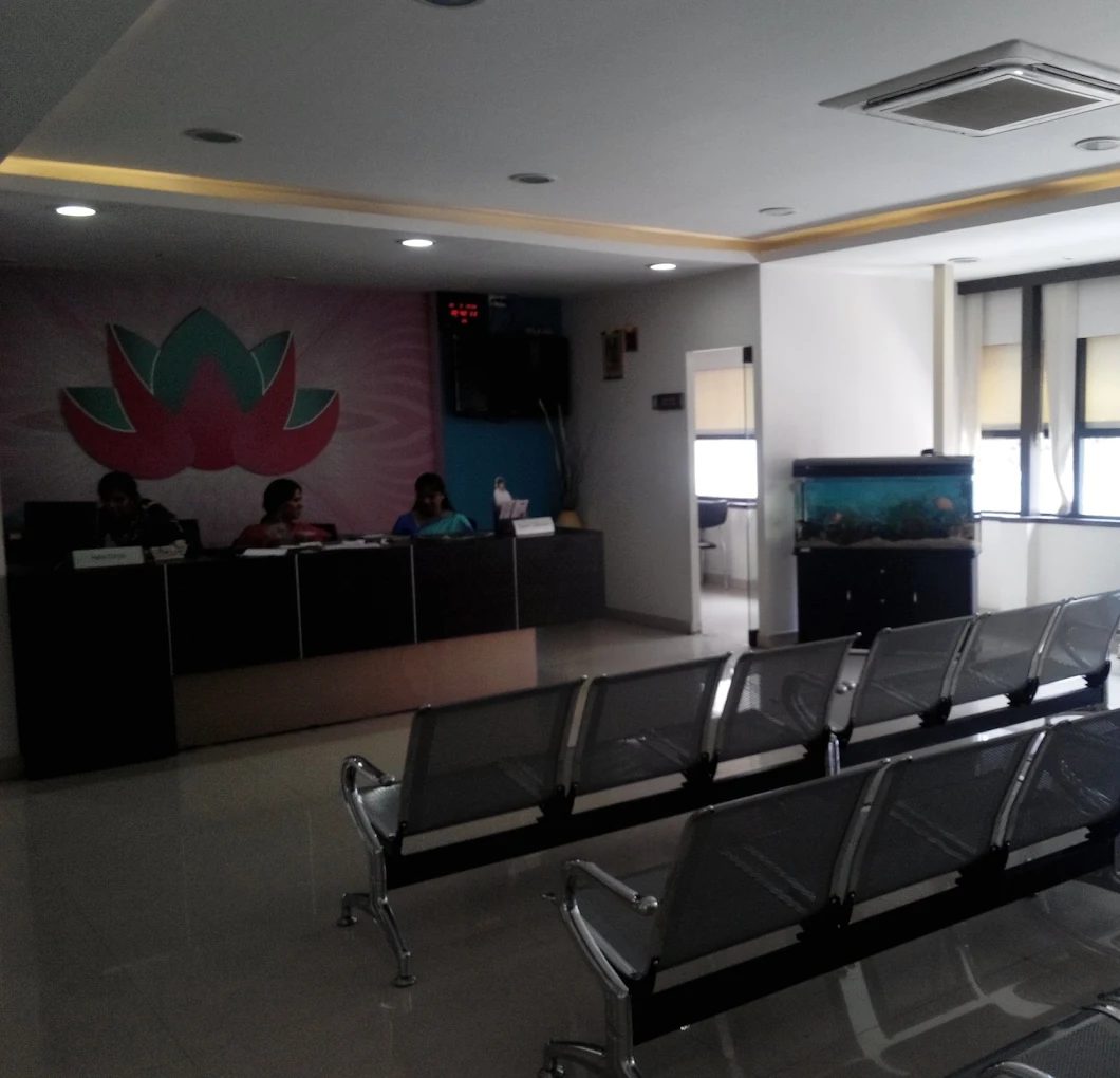 Reception
 Waiting area
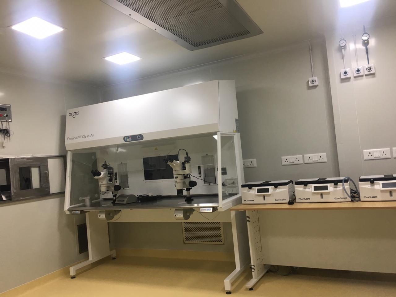 IVF lab
This page explains each test in general terms so you know what to expect. It is not medical advice: your doctor decides which tests you need and what your results mean. Reference ranges differ between laboratories. Updated 4 November 2026.