
Fetal echocardiogram (baby heart scan)
Quick answer: A fetal echocardiogram is a detailed ultrasound of the unborn baby's heart that looks at its chambers, valves, vessels and rhythm. It is usually done around 18 to 24 weeks when there is a higher chance of a heart problem, such as a family history, diabetes, IVF pregnancy or a finding on another scan.
- SampleImaging (no sample)
- TakesAbout 30 minutes to an hour
- Before the testNo special preparation is usually needed.
- In fertility careSome doctors recommend a fetal echo in IVF and ICSI pregnancies, as some studies show a slightly higher rate of heart defects.
How the test is done
- 1You lie on your back with your belly uncovered
- 2Warm gel is spread on your belly and the probe is moved over it
- 3The sonographer looks at the baby's heart chambers, valves, vessels and rhythm
- 4You may be asked to turn slightly if the baby's position makes the heart hard to see
What your results can mean
Reading your report
Measurements are compared with what is expected for the stage of pregnancy; your doctor will explain what your report means for you and your baby.
If the result is outside the usual range
A fetal echo can show differences in how the baby's heart chambers, valves or major vessels have formed, or an unusual heart rhythm. Some small heart problems cannot be seen before birth, and a normal scan lowers but does not remove the chance of a heart condition.
Who usually has this test
Pregnant women with a family history of heart defects, diabetes, an IVF pregnancy, certain medicines, or a heart concern seen on another scan.
Risks and aftercare
It uses sound waves, not radiation, and is considered safe for you and the baby; pressure on a full belly may feel uncomfortable, and there is no aftercare.
Names and options
Also called: fetal echo, foetal echocardiography, baby heart scan
Listed by the hospital as
- DOPPLER FOETAL ECHOCARDIOGRAM
Questions about this test
When is a fetal echo done?
It is usually done around 18 to 24 weeks of pregnancy, when the baby's heart is large enough to see in detail. Sometimes it is repeated later.
Is a fetal echo needed after IVF?
Some doctors recommend it in IVF and ICSI pregnancies because some studies show a slightly higher rate of heart defects. Your doctor will advise based on your situation.
How is a fetal echo different from an anomaly scan?
The anomaly scan checks the whole baby, including a basic heart check. A fetal echo spends much longer on the heart alone, looking at its structure and rhythm in more detail.
Related tests
Tamara Hospital & IVF Centre, Rajajinagar
34/3, 10th Cross Road, opp. Vardhaka Sardar Patel School, 1st N Block, 2nd Stage, Rajajinagar, Bengaluru 560010
The hospital is open 24 hours. Call before you come to check the timing for your scan or sample collection.
Get directions ↗IVF Pulse fertility care at Tamara Hospital →
 Reception
 Waiting area
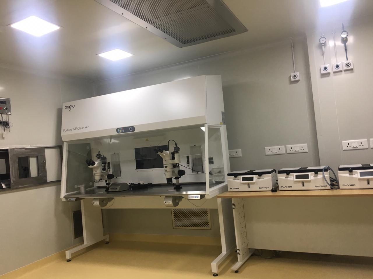 IVF lab
This page explains each test in general terms so you know what to expect. It is not medical advice: your doctor decides which tests you need and what your results mean. Reference ranges differ between laboratories. Updated 4 November 2026.