
Joint and tendon ultrasound
Quick answer: A joint and tendon ultrasound looks at the muscles, tendons, ligaments and fluid around a joint, such as a tendon tear or swelling. Doctors order it for joint or tendon pain, swelling or injury, for example a painful shoulder, heel or Achilles tendon.
- SampleImaging (no sample)
- TakesAbout 15 to 30 minutes
- Before the testNo special preparation is usually needed.
How the test is done
- 1You uncover the joint or area being checked
- 2Gel is spread on the skin over the joint or tendon
- 3The probe is moved over the area while you hold still
- 4You may be asked to move the joint so the tendon can be watched while it moves
What your results can mean
Reading your report
A sonologist or radiologist reports on the scan, and your doctor will explain what the findings mean for you.
If the result is outside the usual range
The scan can show a torn or swollen tendon, fluid in a joint or bursa, a ligament injury, a cyst such as a ganglion, or inflammation around a joint. Deep structures inside the joint may need an MRI.
Who usually has this test
People with shoulder, elbow, wrist, knee, heel or ankle pain, a tendon injury, or a lump near a joint.
Risks and aftercare
There is no radiation; moving a painful joint during the scan may be uncomfortable, and there is no aftercare.
Names and options
Also called: musculoskeletal ultrasound, MSK ultrasound, shoulder ultrasound, knee ultrasound, Achilles tendon scan
Options
- Ankle (Achilles tendon)
- Heel
- Hip joint
- Knee joint
- Shoulder
Listed by the hospital as
- ULTRASOUND SCAN ANKLE (ACHILLES TENDON)
- ULTRASOUND SCAN HEEL
- ULTRASOUND SCAN HIP JOINT
- ULTRASOUND SCAN KNEE JOINT
- ULTRASOUND SCAN SHOULDER
Questions about this test
Can a joint ultrasound show a rotator cuff tear?
Yes, ultrasound is a common way to check the rotator cuff tendons in the shoulder. It can show tears and inflammation.
Why do I need to move my joint during the scan?
Moving the joint lets the sonographer watch how the tendon glides and whether it catches. Some problems only show during movement.
Is an ultrasound or MRI better for a joint injury?
Ultrasound is good for tendons and structures near the surface, and lets the joint be checked while moving. MRI shows deeper parts of a joint, such as the knee cartilage, in more detail.
Related tests
Tamara Hospital & IVF Centre, Rajajinagar
34/3, 10th Cross Road, opp. Vardhaka Sardar Patel School, 1st N Block, 2nd Stage, Rajajinagar, Bengaluru 560010
The hospital is open 24 hours. Call before you come to check the timing for your scan or sample collection.
Get directions ↗IVF Pulse fertility care at Tamara Hospital →
 Reception
 Waiting area
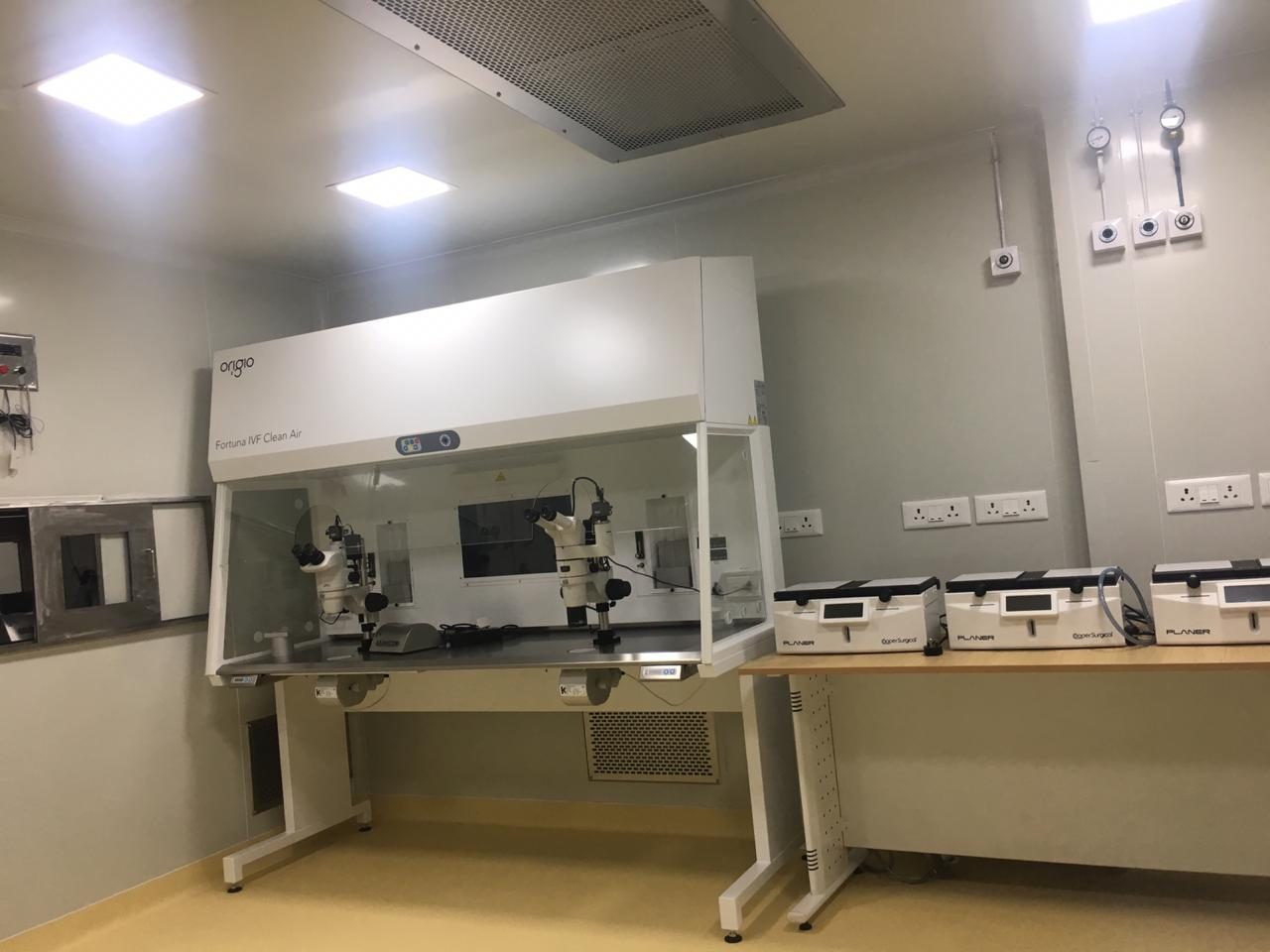 IVF lab
This page explains each test in general terms so you know what to expect. It is not medical advice: your doctor decides which tests you need and what your results mean. Reference ranges differ between laboratories. Updated 4 November 2026.