
Heel bone (calcaneum) X-ray
Quick answer: A heel X-ray shows the heel bone (calcaneum) and can show fractures and bony growths such as heel spurs. Doctors usually order it for heel pain, a fall onto the feet, or swelling around the heel.
- SampleImaging (no sample)
- TakesAbout 5 to 10 minutes
- Before the testNo special preparation is usually needed, but remove metal items from the area and tell staff if you might be pregnant.
How the test is done
- 1You remove your shoe and sock
- 2You lie or sit with the side of your foot resting on the X-ray plate
- 3For a second view your foot is flexed upwards so the heel is seen from below
- 4You hold the foot still briefly while each picture is taken
What your results can mean
Reading your report
A radiologist reports on the images, and your doctor will explain what the findings mean for you.
If the result is outside the usual range
A heel X-ray can show a fracture, often after a fall from a height, a bony spur on the underside or back of the heel, or other bone changes. Plantar fasciitis is a soft-tissue problem and may not show, even when it is causing the pain.
Who usually has this test
People with heel pain on walking, especially first thing in the morning, or anyone who has landed hard on their feet.
Risks and aftercare
Nothing is felt and no aftercare is needed; the radiation is low, but mention it to staff if you could be pregnant.
Names and options
Also called: calcaneum X-ray, calcaneus X-ray, heel X-ray, heel spur X-ray
Options
- Heel from below (axial view)
- Axial view, both sides
- Axial and side views
- Axial and side views, both sides
- Side view (lateral)
- Side view, both sides
- Front view (AP)
- Front and side views (AP and lateral)
Listed by the hospital as
- CALCANEUM AXIAL DIGITAL X-RAY
- CALCANEUM AXIAL (BOTH) DIGITAL X-RAY
- CALCANEUM AXIAL & LAT DIGITAL X-RAY
- CALCANEUM AXIAL & LAT (BOTH) DIGITAL X-RAY
- CALCANEUM LATERAL DIGITAL X-RAY
- CALCANEUM LATERAL (BOTH) DIGITAL X-RAY
- HEEL AP DIGITAL X-RAY
- HEEL AP, LATERAL DIGITAL X-RAY
- HEEL LATERAL DIGITAL X-RAY
Questions about this test
Does a heel spur on X-ray mean it is causing my pain?
Not always. Many people have heel spurs without any pain, and heel pain is often from the plantar fascia, a band of tissue under the foot. Your doctor will put the X-ray together with your symptoms and examination.
Can a heel X-ray show plantar fasciitis?
Plantar fasciitis is usually diagnosed from symptoms and examination. An X-ray mainly rules out a fracture or other bone cause, while an ultrasound can show thickening of the fascia.
Why is a heel X-ray taken from below?
The view from below, called an axial view, shows the width of the heel bone. It helps find fractures that can be missed on the side view alone.
Related tests
Tamara Hospital & IVF Centre, Rajajinagar
34/3, 10th Cross Road, opp. Vardhaka Sardar Patel School, 1st N Block, 2nd Stage, Rajajinagar, Bengaluru 560010
The hospital is open 24 hours. Call before you come to check the timing for your scan or sample collection.
Get directions ↗IVF Pulse fertility care at Tamara Hospital →
 Reception
 Waiting area
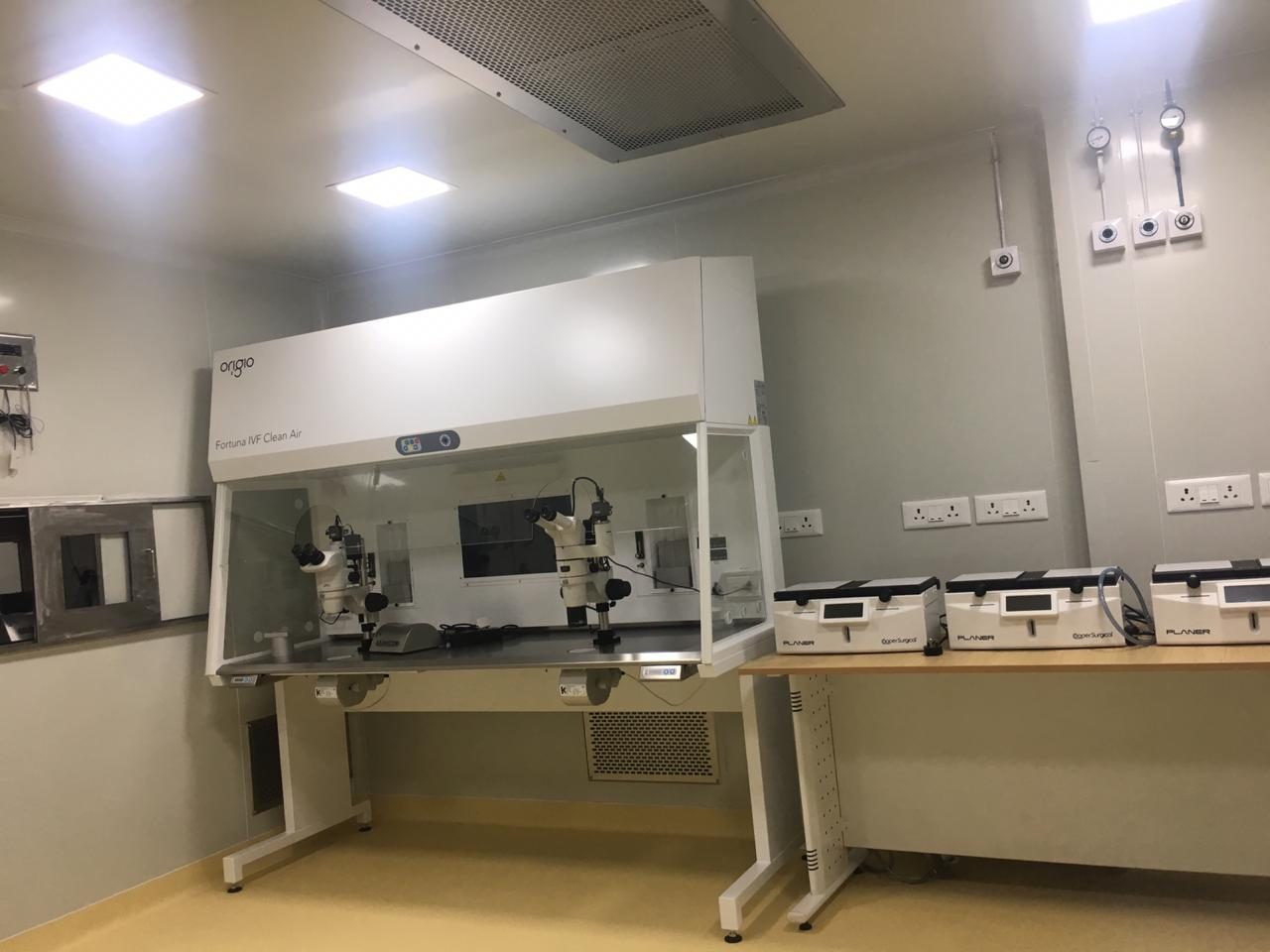 IVF lab
This page explains each test in general terms so you know what to expect. It is not medical advice: your doctor decides which tests you need and what your results mean. Reference ranges differ between laboratories. Updated 4 November 2026.