
Foot X-ray
Quick answer: A foot X-ray shows the bones of the foot and toes and can show fractures, joint changes and bony growths. Doctors usually order it after a foot injury, or for foot pain, swelling or deformity.
- SampleImaging (no sample)
- TakesAbout 10 minutes
- Before the testNo special preparation is usually needed, but remove metal items from the area and tell staff if you might be pregnant.
How the test is done
- 1You remove your shoes, socks and toe rings
- 2You sit on the table with your knee bent and the sole of your foot flat on the plate
- 3For a turned view the foot is tilted inwards slightly
- 4A standing view may be taken to show the foot under body weight
What your results can mean
Reading your report
A radiologist reports on the images, and your doctor will explain what the findings mean for you.
If the result is outside the usual range
A foot X-ray can show a fracture, including stress fractures in later stages, joint changes from arthritis or gout, a bunion, flat feet on standing views, or bone infection in people with diabetic foot problems.
Who usually has this test
People with a foot injury, pain under or across the foot, a bunion, or people with diabetes and a foot ulcer or swelling.
Risks and aftercare
Putting weight on a sore foot for a standing view may hurt briefly, so tell staff if you cannot; radiation to the foot is low, and mention any chance of pregnancy.
Names and options
Also called: X-ray foot
Options
- Front view (AP)
- Front and side views (AP and lateral)
- Front and side views, both sides
- Front and angled views (AP and oblique)
- Front and angled views, both sides
Listed by the hospital as
- FOOT AP DIGITAL X-RAY
- FOOT AP & LATERAL DIGITAL X-RAY
- FOOT AP & LATERAL (BOTH) DIGITAL X-RAY
- FOOT AP & OBLIQUE DIGITAL X-RAY
- FOOT AP & OBLIQUE (BOTH) DIGITAL X-RAY
Questions about this test
Why is a foot X-ray taken standing?
A standing view shows how the bones line up when they carry your weight. This matters for flat feet, bunions and some joint problems that look different when the foot is resting.
Can a foot X-ray show gout?
In early gout the X-ray is often normal. In long-standing gout it can show typical bone erosions near the big toe joint, and a blood uric acid test is also used.
Can an X-ray show a fracture from overuse in the foot?
Stress fractures often do not show in the first two weeks. As they heal, new bone forms around the crack and becomes visible on a repeat X-ray.
Related tests
Tamara Hospital & IVF Centre, Rajajinagar
34/3, 10th Cross Road, opp. Vardhaka Sardar Patel School, 1st N Block, 2nd Stage, Rajajinagar, Bengaluru 560010
The hospital is open 24 hours. Call before you come to check the timing for your scan or sample collection.
Get directions ↗IVF Pulse fertility care at Tamara Hospital →
 Reception
 Waiting area
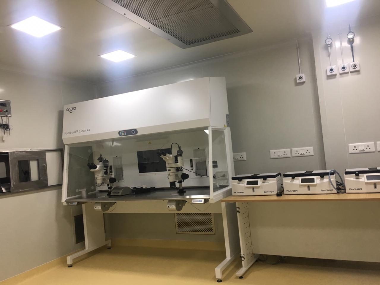 IVF lab
This page explains each test in general terms so you know what to expect. It is not medical advice: your doctor decides which tests you need and what your results mean. Reference ranges differ between laboratories. Updated 4 November 2026.